
Understanding cell division, mitosis, is crucial for comprehending growth and development in living organisms. Onion root tips provide an accessible model for observing the dynamic stages of plant cell mitosis. This section outlines foundational aspects of studying this vital biological process effectively.
Significance of Cell Division in Plants
Cell division, specifically mitosis, is unequivocally fundamental to the life cycle and overall success of plants. It underpins virtually all aspects of their growth, development, and repair mechanisms. Through precisely regulated mitotic processes, plants are able to increase their biomass, forming complex structures such as extensive root systems, sturdy stems, and photosynthetically active leaves. This continuous proliferation of cells originates primarily from specialized regions known as meristems, found at the tips of roots and shoots, as well as in the cambium. Without consistent cell division, a plant could not develop from a tiny seed into a mature organism capable of reproduction. Moreover, mitosis is essential for tissue repair following injury, allowing plants to regenerate damaged parts or recover from environmental stressors. It also plays a critical role in asexual reproduction, where a single parent plant can produce genetically identical offspring. The ability to generate new cells ensures the ongoing vitality and adaptive capacity of plant species across diverse ecosystems, making it a cornerstone of botany and agricultural science. Observing these processes firsthand, such as in onion root tips, provides invaluable insight into these biological imperatives. This constant renewal and expansion highlight the profound importance of mitotic activity in sustaining plant life, from microscopic cellular changes to macroscopic ecological impact.
Why Onion Root Tips are Ideal for Mitosis Study
Onion root tips are exceptionally well-suited for the study of mitosis in a laboratory setting, making them a staple in biology education. Several key characteristics contribute to their ideal nature. Primarily, the apical meristem, located at the very tip of the onion root, is a region of intense cellular proliferation. This means a high percentage of cells within a prepared slide will be actively undergoing various stages of mitosis, providing ample opportunities for observation of prophase, metaphase, anaphase, and telophase. Unlike some other plant tissues, onion root tip cells possess relatively large chromosomes that are distinct and easily visible under a light microscope, especially after proper staining. Furthermore, the diploid chromosome number for onions (2n=16) is manageable, simplifying the identification and tracking of individual chromosomes through the division process; Their ease of cultivation is another significant advantage; onions can be readily sprouted in water, producing a continuous supply of healthy root tips. The preparation of slides is also straightforward, involving simple fixation, maceration, and squashing techniques that yield a single layer of cells, ideal for clear microscopic examination. These combined attributes make the onion root tip an invaluable model for hands-on learning about the fundamental process of cell division.
Lab Objectives and Hypotheses
This lab aims to identify and describe the distinct stages of mitosis within onion root tip cells. We hypothesize interphase will be the most frequently observed stage, reflecting its longer duration in the cell cycle.
Goals for Identifying Mitotic Stages
The primary goal of this laboratory exercise is to cultivate a comprehensive understanding of the eukaryotic cell cycle, specifically focusing on the process of mitosis as observed in the meristematic cells of onion root tips. Students are expected to develop proficiency in microscopically identifying and distinguishing between the various phases of mitosis: prophase, metaphase, anaphase, and telophase. A crucial objective involves recognizing the characteristic morphological changes within the nucleus and cytoplasm that define each stage, such as chromosome condensation, nuclear envelope breakdown, spindle fiber formation, chromosome alignment at the metaphase plate, sister chromatid separation, and cytokinesis. Furthermore, an important aim is to differentiate these active mitotic stages from the non-dividing interphase cells, which typically comprise the majority of observed cells and represent periods of growth and DNA replication. Through careful observation of prepared slides, participants will enhance their practical microscopy skills, including proper focusing, illumination, and stage manipulation to locate and analyze cells undergoing division. Ultimately, these identification skills are foundational for subsequent quantitative analyses, such as calculating the mitotic index, and for appreciating the fundamental mechanisms of growth and repair in multicellular organisms. The ability to accurately categorize cells into their respective stages is paramount for any successful investigation into cell proliferation rates or the effects of various agents on the cell cycle.

Expected Observations of Cell Cycle Phases

Expected microscopic observations will reveal distinct characteristics for each cell cycle phase in onion root tip cells. Most cells will be in interphase, showing a prominent nucleus with dispersed chromatin, lacking visibly condensed chromosomes, indicating active growth. During prophase, chromosomes condense, becoming visible threads, and the nuclear envelope begins to disappear. Metaphase is identified by chromosomes precisely aligned at the cell’s equatorial plate, forming a distinct line. In anaphase, sister chromatids separate and move towards opposite poles, often appearing characteristically V-shaped. Telophase features chromosomes clustered at each pole, decondensing, with new nuclear envelopes reforming. Concurrently, a cell plate forms centrally, leading to cytokinesis, the complete division of cytoplasm into two genetically identical daughter cells. These crucial morphological changes are vital for accurate identification and analysis.

Materials and Experimental Procedure
This section outlines the essential materials and detailed steps for the onion root tip mitosis lab. It covers cultivating onion roots, preparing microscopic slides, and setting up the microscope for optimal viewing and identification of various mitotic stages.
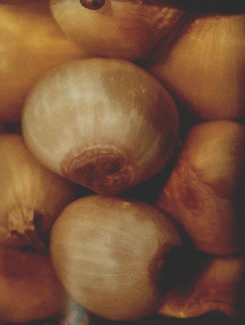
Growing Onion Roots For Mitosis
To successfully observe mitosis in onion root tips, the initial step involves carefully cultivating fresh, actively growing roots. Begin by selecting a healthy, firm onion bulb, preferably one that has not started sprouting already. Remove any dry, outer layers to expose the base. Position the onion bulb over a glass jar or beaker filled with water, ensuring that only the bottom portion of the bulb, where the roots will emerge, is submerged in the water. Toothpicks can be inserted around the onion’s circumference to help suspend it securely above the water, preventing the entire bulb from being immersed. The water level should be maintained consistently, just touching the base of the onion. Place the setup in a warm, well-lit area, but avoid direct sunlight which might inhibit growth or promote algal development. Replace the water every one to two days to prevent stagnation and bacterial growth, which could harm the developing roots. Within approximately three to five days, new roots, typically about 2-3 centimeters long, will begin to grow from the base of the onion. These newly developed root tips contain the apical meristem, a region of rapid cell division, making them ideal specimens for studying the various stages of mitosis. Timely harvesting of these fresh, vigorous roots is critical for obtaining high-quality samples for subsequent slide preparation and microscopic analysis, ensuring a clear observation of the cell cycle phases.
Preparation of Onion Root Tip Slides
Once fresh onion root tips are grown, slide preparation begins with fixation. Root tips, 1-2 cm long, are immersed in a fixative like Carnoy’s fluid for 12-24 hours. This essential step halts cell division and preserves cellular structures, preventing degradation and maintaining chromosome integrity for accurate observation of chromosomal activities under a microscope.
Next, maceration softens cell walls, allowing tissue to spread into a single, flat layer. Tips are then gently heated in 1M HCl at 60°C for a few minutes to dissolve the pectin-rich middle lamella. After thorough rinsing with distilled water to neutralize the acid, the roots are stained. Acetocarmine or aceto-orcein are common choices, binding to chromatin and coloring chromosomes (red or purple) over 15-30 minutes.
For the final slide, the meristematic tip (1-2 mm) is placed on a clean slide with fresh stain. A coverslip is lowered, and gentle, even pressure is applied to squash the tissue, creating a single cell layer that is crucial for clear cell observation. Sealing the coverslip with nail polish prevents dehydration, ensuring the slide remains viable for extended, optimal visualization of mitotic stages.
Microscope Setup and Viewing Technique
Proper microscope setup is paramount for successful observation of onion root tip mitosis; Begin by placing the prepared slide securely on the microscope stage, ensuring the coverslip faces upwards. Start with the lowest power objective lens (typically 4x or 10x) to locate the meristematic region, which is the area of active cell division just behind the root cap. Use the coarse adjustment knob to bring the image into initial focus, then refine with the fine adjustment knob.
Once the meristematic zone is identified, rotate to a higher power objective lens (e.g., 40x). Recalibrate the focus using only the fine adjustment knob, as the coarse adjustment can cause the objective lens to crash into the slide at high magnifications. Adjust the diaphragm or iris to control the light intensity and contrast, providing the clearest view of cellular structures and chromosomes. A systematic scanning technique, moving the stage methodically across the meristematic region, is essential to locate various cells undergoing different stages of mitosis. This careful approach ensures no dividing cells are overlooked, maximizing data collection for your lab report.

Observations: Mitotic Stages and Cell Counts
This section presents visual observations from onion root tip slides. We identify and describe distinct mitotic stages. Data collection involves systematically counting cells within each cell cycle phase, forming the basis for quantitative analysis in this lab.
Mitosis Lab Onion Root Tip Images and Photos
Data Collection: Number of Cells in Each Phase
Data collection is a critical step in the onion root tip mitosis lab, enabling quantitative analysis of the cell cycle. After meticulously preparing the slides and setting up the microscope, students systematically scan the meristematic region, where cell division is most active. The objective is to count observed cells in each distinct phase: interphase, prophase, metaphase, anaphase, and telophase. This systematic counting involves traversing the field of view in a structured manner to avoid recounting or missing sections. A tally system or digital counter is used to record the frequency of each phase encountered. Accurate identification, aided by visual resources like docsity.com or an “Onion Root Tip Cell Cycle Lab Worksheet” (studylib.net), is paramount for reliable results. These raw numbers collected represent the prevalence of cells in different stages, indirectly reflecting the relative duration of each phase. This quantitative data forms the foundation for subsequent calculations, such as the mitotic index, providing empirical evidence. Without cell counts, comprehensive analysis of plant growth and cellular activity would be impossible, making this step indispensable for any mitosis lab report answer key;
Calculating the Mitotic Index
The mitotic index is a fundamental quantitative measure in the onion root tip mitosis lab, reflecting the proportion of cells actively undergoing division. This crucial index is determined by dividing the total count of cells observed in mitosis (prophase, metaphase, anaphase, telaphase) by the grand total of all cells counted within the microscopic field, including interphase cells. The resulting ratio is multiplied by 100 to express the mitotic index as a percentage. This provides direct insight into the cell proliferation rate of the meristematic tissue. A higher mitotic index signifies more rapid growth, a characteristic expected in the onion root tip’s apical region. Conversely, a lower index suggests a slower rate of cell division. This calculation is essential for interpreting experimental data on plant growth and overall cellular activity, offering a clear summary of the observed cell cycle dynamics.

Detailed Analysis of Cell Cycle Phases
This section delves into the intricate characteristics of each phase within the cell cycle, as observed in onion root tip cells. We will meticulously examine the distinct morphological changes that define interphase and the subsequent stages of mitosis, providing a comprehensive understanding.
Interphase and its Sub-phases
Interphase represents the longest duration of the cell cycle, a period of intense cellular activity and growth rather than dormancy. In onion root tip cells, observing interphase under a microscope reveals a well-defined nucleus with visible nucleoli and chromatin that appears as an uncoiled, diffuse mass, lacking distinct chromosomes. This appearance is characteristic of cells preparing for division or carrying out normal metabolic functions, as often detailed in “Onion Root Tip Cell Cycle Lab Worksheet” and similar study materials.
This critical preparatory stage is subdivided into three distinct phases: G1, S, and G2. During the G1 (Gap 1) phase, the cell undergoes significant growth, synthesizing proteins and organelles essential for its function; Following G1 is the S (Synthesis) phase, a crucial period where the cell’s DNA is replicated, ensuring that each daughter cell receives a complete set of genetic material. Finally, the G2 (Gap 2) phase involves further growth and the synthesis of additional proteins and structures, specifically those required for the upcoming mitotic division, such as microtubules. Identifying these sub-phases microscopically is often challenging without specialized staining or techniques, but their collective impact is evident in the cell’s readiness for mitosis.
Prophase Onion Root Tip Cells Mitosis
Prophase marks the initial and often the longest stage of mitosis in onion root tip cells, a period of significant reorganization within the nucleus. As observed in various “Mitosis Lab Onion Root Tip Images and Photos,” the most striking change is the condensation of chromatin. The diffuse genetic material, characteristic of interphase, begins to coil tightly, becoming distinctly visible as individual, thread-like chromosomes under a light microscope. Each chromosome at this stage consists of two identical sister chromatids, joined at a centromere, resulting from the DNA replication during the S-phase of interphase.
Simultaneously, the nucleolus, a dense structure within the nucleus, gradually disappears. This indicates a cessation of ribosomal RNA synthesis; Concurrently, the nuclear envelope, which encloses the genetic material, starts to fragment and dissolve, allowing the forming spindle microtubules to gain access to the chromosomes. Although not always clearly visible in all preparations of onion root tips, the initiation of spindle fiber formation from the centrosomes (though plant cells lack centrioles, they organize microtubules) is a critical event; These cellular transformations prepare the chromosomes for their precise segregation in subsequent mitotic stages.
Metaphase, Anaphase, and Telophase Characteristics
Following prophase, Metaphase in onion root tip cells is defined by the precise alignment of condensed chromosomes along the cell’s equatorial plate. Spindle fibers attach to the centromeres of each sister chromatid. This critical alignment ensures equal genetic distribution to daughter cells, a key observation in “Onion Root Tip Analysis Lab Stages of Mitosis Study notes.”
Next, Anaphase begins with the synchronous separation of sister chromatids. Centromeres divide, and individual chromosomes are pulled towards opposite poles by shortening spindle fibers. This active migration results in two distinct groups of chromosomes moving apart, often elongating the cell. This crucial segregation ensures genetic identity for new cells.
Telophase concludes nuclear division. At the poles, chromosomes decondense into chromatin. New nuclear envelopes reform around each set of chromosomes, and nucleoli reappear. Concurrently, cytokinesis starts with cell plate formation at the former metaphase plate, ultimately dividing cytoplasm and yielding two distinct daughter cells, completing mitosis in the onion root tip.
Onion Root Tip Analysis Lab Stages of Mitosis Study Notes
For effective observation in an onion root tip mitosis lab, these study notes from resources like Docsity’s “Onion Root Tip Analysis Lab Stages of Mitosis Study notes” are invaluable. Focus on clear identification cues for each phase under the microscope. During Interphase, look for a large, intact nucleus with no distinct chromosomes. It’s the longest phase, so most cells will appear here. For Prophase, observe the condensing chromosomes becoming visible as thread-like structures, and the nuclear envelope beginning to disappear. The cell might appear denser.
Discussion of Results and Potential Errors
This section analyzes observed data, comparing it to expected mitotic patterns. We’ll examine the calculated mitotic index and identify sources of experimental variation. Potential errors include slide preparation issues or misidentification of cell phases, impacting result accuracy.
Interpreting the Mitotic Index
The mitotic index (MI) is a crucial quantitative measure in onion root tip analysis, representing the proportion of cells actively undergoing mitosis relative to the total cell population. Calculated by dividing the number of mitotic cells by the total number of observed cells, it directly reflects the tissue’s growth rate and proliferative activity. A high mitotic index signifies robust cell division, characteristic of the rapidly growing meristematic zone in root tips where new cells are constantly being produced for elongation. Conversely, a lower MI suggests reduced cellular proliferation, which could be indicative of slower growth, cellular stress, or the influence of environmental factors inhibiting the cell cycle. This index also provides insight into the relative duration of mitosis within the entire cell cycle; a shorter mitotic phase in comparison to interphase will result in a lower MI, even if the cell cycle is active. Interpreting the mitotic index allows for comprehensive evaluation of the experiment’s findings, facilitating the identification of cellular responses to various conditions, such as the impact of chemical treatments or varying temperatures. It’s essential for validating hypotheses and drawing sound conclusions about cell cycle dynamics, forming a fundamental analytical component of lab reports and their answer keys.

Onion Root Tip Mitosis Lab Report Answer Key
This section provides the essential components for a correct onion root tip mitosis lab report, serving as a comprehensive answer key. Students are expected to accurately identify all mitotic phases—interphase, prophase, metaphase, anaphase, and telophase—based on distinct cellular morphology from their microscope observations, often referenced in “Onion Root Tip Analysis Lab Stages of Mitosis Study notes.” A critical finding is the overwhelming presence of interphase cells, signifying the majority of a cell’s life cycle. Data collection must include precise counts of cells in each phase across multiple fields of view for statistical validity, which then informs the calculation of the mitotic index. A healthy onion root tip typically exhibits a mitotic index reflecting active, rapid growth, consistent with plant meristematic tissue. Discrepancies in cell counts or the mitotic index may indicate experimental issues, such as errors in slide preparation or observation, or external factors affecting cell division. The answer key emphasizes understanding how these microscopic observations directly illustrate the fundamental process of growth and development in plants, a core concept frequently assessed in “Onion Root Tip Mitosis Lab Worksheets” for a complete understanding.